How many ultrasounds do you get during pregnancy? While most low-risk pregnancies involve two routine scans, additional ultrasounds may be recommended to monitor growth, positioning, or potential complications. Learn when you may need more ultrasounds and how to prepare for your ultrasound appointment.
Table of Contents
How Does An Ultrasound Scan In Pregnancy Work?
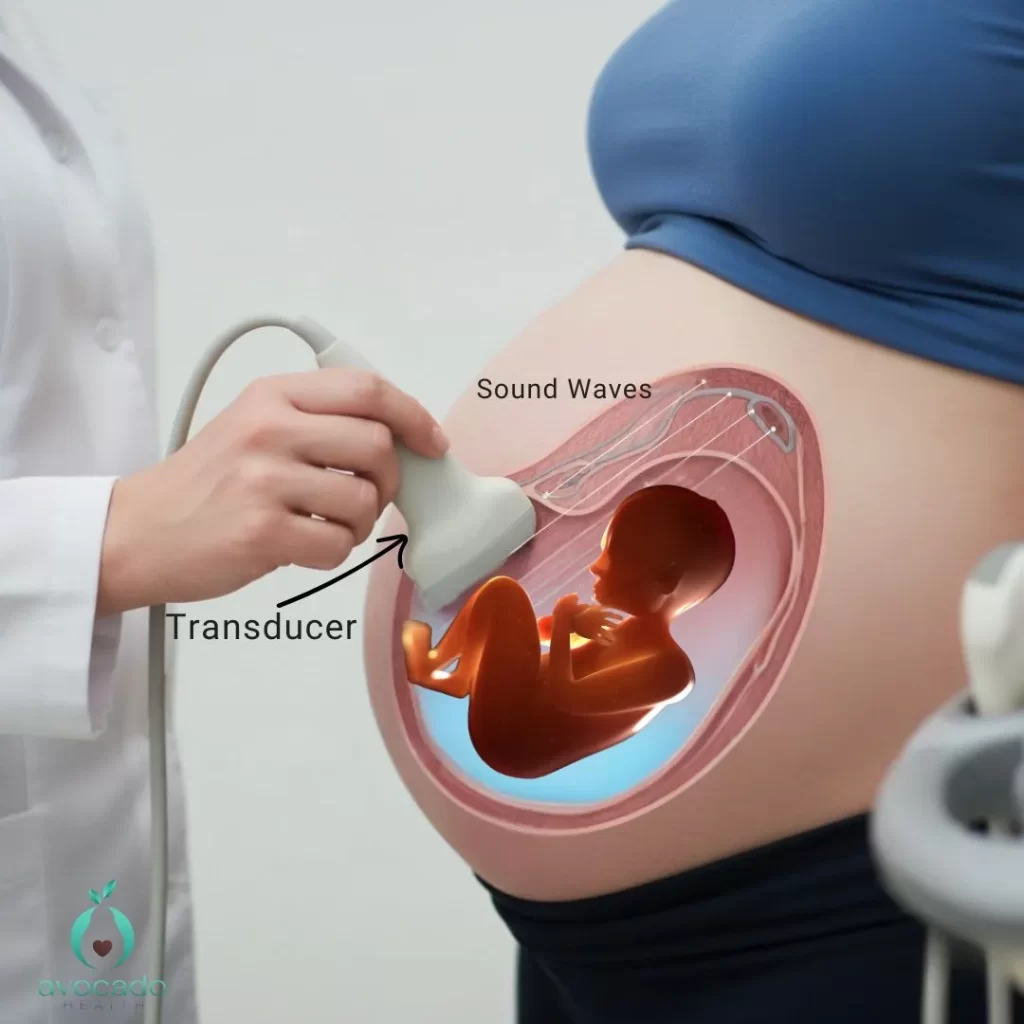
An ultrasound scan during pregnancy uses high-frequency sound waves to create images of the developing baby inside the womb.
A device called a transducer emits these sound waves, which travel through the mother’s abdomen or vagina and bounce off the baby and surrounding tissues. The echoes return to the transducer and are converted into real-time images displayed on a monitor.
How Many Ultrasounds Do You Get During Pregnancy and what to expect

The number of ultrasounds during pregnancy varies but typically includes at least two. Here’s a quick overview:
- Typical Ultrasound Count: Most women have at least two ultrasounds during a healthy, low-risk pregnancy, but several ultrasounds may be performed throughout pregnancy, especially if there are medical concerns.
- Standard Ultrasounds:1- First Trimester Ultrasound: Usually done between 8 and 12 weeks (most commonly 10-13 weeks). Also called the viability and dating ultrasound. Key purposes:1
- Confirm pregnancyCheck fetal heartbeatEstimate due dateDetermine baby’s gestational age for prenatal care planning
- Performed between 18-22 weeks
- Examines baby’s organs and development
- Measures baby’s growth and size
- Assesses amount of amniotic fluid
- Checks baby’s sex (if desired and position allows)
- Additional Ultrasounds May Be Needed If:
- Concerns arise about baby’s growth (e.g., intrauterine growth restriction).
- Medical conditions like gestational diabetes or high blood pressure are present.
- Monitoring of amniotic fluid levels or placenta position (e.g., placenta previa) is necessary.
- More ultrasounds may be required in a high risk pregnancy, for expecting twins, or to monitor baby’s growth and development.
- Third Trimester Ultrasound:
- Sometimes done, when indicated, between 28-40 weeks to check baby’s position (head down or breech), size, and well-being before labor.2
- Around 36 weeks of pregnancy, your provider will do a quick ultrasound to check the baby’s position for delivery, which helps plan for a safe vaginal delivery and monitor any late pregnancy concerns.
- Multiple Pregnancies: If you are expecting twins, more frequent ultrasounds are needed to closely track each baby’s growth and development.
Overall, your healthcare provider will tailor the ultrasound schedule based on your specific needs to support a healthy pregnancy.
Ultrasounds are typically performed by an ultrasound technician or OB-GYN and should only be done when there is a valid medical indication.
Is 12 Week Ultrasound Internal Or External?
At 12 weeks, the ultrasound is usually performed abdominally, but a transvaginal ultrasound may be used if clearer images are needed.
This 12-week scan, sometimes called an early ultrasound, helps to:3
- Confirm ongoing pregnancy and fetal development
- Check the embryo’s location and size
- Assess fetal viability
A transvaginal ultrasound involves inserting a small probe into the vagina, allowing the ultrasound technician to get closer to the uterus and developing baby. This method is especially useful during the first trimester when the baby is still very small and abdominal ultrasounds may not provide sufficient detail.
Is It A Boy Or Girl Scan At 12 Weeks?
Many expectant parents are eager to find out their baby’s sex as early as possible. Here’s what you need to know about the 12-week ultrasound and baby’s sex:
- Primary Focus: The 12-week ultrasound, often called the dating or nuchal translucency scan, mainly assesses the baby’s development and screens for genetic conditions.
- Sex Determination: Sometimes, this scan may offer a glimpse of the baby’s sex, but it is challenging and not always accurate due to the baby’s small size and position.
Why No Ultrasound Until 8 Weeks?

In many pregnancies, healthcare providers recommend waiting until around 8 weeks to perform the first ultrasound. This timing is intentional and based on several important reasons: 4
- Early Development Stage: Before 8 weeks, the embryo is extremely small and still developing rapidly. It can be challenging to get clear and accurate images that provide meaningful information about the pregnancy.
- Detecting Fetal Heartbeat: Around 6 to 7 weeks, the baby’s heartbeat may just begin to be detectable via ultrasound, but it is more reliably seen by 8 weeks. Performing an ultrasound too early might result in inconclusive findings, leading to unnecessary anxiety.
- Reducing False Alarms: Early ultrasounds may sometimes show what appears to be an empty gestational sac or no heartbeat, which can be normal at very early stages but may cause confusion or concern. Waiting until 8 weeks helps ensure more definitive and reassuring results.
- Minimizing Repeat Ultrasounds: By scheduling the first ultrasound around 8 weeks, healthcare providers can reduce the need for multiple repeat ultrasounds due to unclear early images, making the process more efficient for both patients and providers.
For women with a medical history of miscarriage or ectopic pregnancy, an earlier ultrasound might be recommended to confirm the pregnancy is developing inside the uterus and progressing normally.
How Many Ultrasounds Do You Get In The Third Trimester?
While many women with low-risk pregnancies may not need any ultrasounds in this final stage, others may require one or more scans to ensure the baby’s health and readiness for birth.
Why Is Ultrasound Important In Third Trimester?

- Assess baby’s size and overall well-being before labor
- Determine baby’s position (head-down, breech, or other)
- Evaluate amniotic fluid levels for potential concerns
- To update ultrasound results and guide interventions for a healthy delivery
Swelling in the legs and feet is common later in pregnancy, and while it’s often normal, sudden or severe swelling should always be discussed with your doctor, especially when paired with other symptoms.
Does Everyone Have A 32 Week Scan?
Not everyone will have a 32-week ultrasound scan as part of their routine prenatal care.
- Who Needs It? A 32-week scan is often recommended for high-risk pregnancies or if earlier scans raised concerns.
- Common High-Risk Factors:
- Gestational diabetes
- High blood pressure
- Carrying twins or multiples
- Signs of intrauterine growth restriction (IUGR)
This scan helps closely monitor fetal well-being and guides care decisions as the pregnancy progresses.
Do You Get An Ultrasound At Every OB Appointment?
It is not typical to have an ultrasound at every OB (obstetrician) appointment during pregnancy. Most standard prenatal visits focus on checking the mother’s health, measuring the baby’s growth through physical examination, and discussing any symptoms or concerns. Ultrasounds are usually scheduled at specific milestones or when medically indicated rather than at every visit.
Can Ultrasounds Miss Birth Defects?
While ultrasounds are a vital tool for monitoring fetal development and detecting many birth defects, they are not foolproof and can sometimes miss certain abnormalities. The ability of an ultrasound to detect birth defects depends on several factors:
- Type of defect: Major structural abnormalities like cleft lip or issues with the heart, brain, or kidneys are often detected during the second trimester anatomy scan.
- Gestational age: Some conditions may develop later in pregnancy and might not be visible early on.
- Equipment quality: Higher-quality ultrasound machines provide clearer images.
- Technician skill: The expertise of the ultrasound technician or healthcare provider affects detection accuracy.
Smaller or less obvious defects may not be visible on ultrasound images. In some cases, specialized imaging techniques are needed for diagnosis.
Can Ultrasound Affect The Fetus?
Ultrasound is considered a safe and non-invasive procedure when performed by trained professionals for medical reasons.
- No Radiation: Ultrasound uses sound waves, not radiation like X-rays, making it safe for monitoring fetal development.
- How It Works: Sound waves bounce off the baby’s tissues and organs, creating echoes that form visual images on a screen.
- Safety: Generally safe with no known risks when used appropriately. Extensive research shows no harmful effects on the fetus from standard diagnostic ultrasounds.
Ultrasounds should be performed only when medically necessary. Non-medical use, such as keepsake videos or photos, is discouraged.
Do Babies Feel Ultrasound Waves?
Ultrasound waves used in prenatal scans are sound waves that don’t cause any pain or discomfort to the baby. The amniotic fluid around them acts like a cushion, shielding the little one from the waves – no need to worry!
What To Eat Before Pregnancy Ultrasound?

While there aren’t any hard-and-fast rules about what to eat or drink before your ultrasound, certain foods and drinks can make the experience more comfortable and even improve the images.
1- Staying Hydrated Is Super Important
Drinking plenty of water before your ultrasound is a must—especially early in your pregnancy. A full bladder helps the ultrasound technician get a clearer view of your developing baby, particularly during early ultrasounds like the first trimester dating ultrasound. Typically, your doctor will advise you to:
- Drink several large glasses of water about an hour before your appointment
- Avoid urinating until after the scan
Following these simple steps can improve image quality and make your ultrasound experience smoother.
2- Avoid Foods That Give You Bloat
Try to steer clear of foods that make you feel bloated or gassy, such as beans, carbonated drinks, and deep-fried foods, before your ultrasound. Too much gas can disrupt sound waves, making it harder for the ultrasound tech to get clear images.
3- Eat a Light, Easy Meal
Eating something light and balanced before the ultrasound helps you feel comfortable and prevents hunger during the procedure. Opt for easy-to-digest foods like:
- Fruit
- Vegetables
- Yogurt
- Whole-grain toast
It’s a good idea to eat something before your ultrasound, especially if you’re far along in your pregnancy.
For later ultrasounds, like the anatomy scan or third-trimester ultrasound, a full bladder isn’t usually necessary, but still drink lots of water to stay comfy.
How To Make Baby Active For Ultrasound?
Ultrasound techs often try to get your baby to move around during the scan to get better images or a clearer view of certain parts of their body. Here are a few tips that might help get your baby moving:
- Have a Little Snack: Eating a small, healthy snack about 30 minutes before your ultrasound can sometimes get your baby going. Try a bit of fruit, fruit juice, or a small piece of chocolate – the sugar might do the trick.
- Change Your Position: Gently changing your position during the scan can sometimes get your baby to shift or move around a bit. For example, rolling from side to side or sitting up for a bit can help.
If changing positions causes discomfort, especially in late pregnancy, gentle movement and posture support can help reduce strain on your lower back.
- Gentle Tapping or Talking: Some parents find that softly tapping on their belly or talking to their baby during the scan can get a reaction. The sonographer might gently apply slight pressure on your abdomen to obtain the clearest possible images of your baby.
- Stay Relaxed: Your baby can pick up on your stress levels, so try to stay calm and relaxed before and during the ultrasound to create a comfy environment for your little one.
- Timing Matters: Babies, like kids, have natural sleep cycles. Scheduling your ultrasound at a time when your baby is usually more active might increase the chances of getting some movement on camera.
Feeling unsure about your ultrasound schedule or what to expect next? Text Avocado Health for instant, expert-backed, personalized guidance throughout your pregnancy.
Conclusion
Most pregnancies include at least two routine ultrasounds, but the exact number can vary based on your health, your baby’s growth, and medical needs.
Frequently Asked Questions
How early can pregnancy show on an ultrasound?
Pregnancy can often be picked up on an ultrasound as early as 4 to 5 weeks after your last period. The ultrasound might show the gestational sac, the first sign of pregnancy, inside the uterus.
Is fasting needed for pregnancy ultrasound?
The answer is usually no, fasting isn’t required before a pregnancy ultrasound.
What’s the difference between 2D, 3D, and 4D ultrasounds?
2D ultrasounds show flat, real-time images of the baby. 3D ultrasounds create three-dimensional still images revealing depth and detail. 4D ultrasounds add real-time movement to 3D images, showing the baby’s body in motion. All types are safe and used for different diagnostic or bonding purposes.
Sources:
- Impact of first-trimester ultrasound on early detection of major fetal anomalies
- Accuracy of antenatal ultrasound in predicting large-for-gestational-age babies
- Role of ultrasound in the evaluation of first-trimester pregnancies in the acute setting
- Can Early First Trimester Ultrasounds Correctly Determine Gestational Age Compared to Ultrasounds Performed Between 7 to 14 Weeks Gestational Age
- Effectiveness of routine third trimester ultrasonography to reduce adverse perinatal outcomes in low risk pregnancy